S1 S2 S3 Pattern
S1 S2 S3 Pattern - Percutaneously inserting one or more bone needles into the sacrum under fluoroscopy and/or ct visual guidance. Web the s 1, s 2, s 3 syndrome may be within normal limits in children but in adults raises the posibility of right ventricular enlargement. Web the data obtained using body surface potential mapping suggest that an anomalous wavefront rightward and superiorly oriented is present in the s1s2s3. Other features of rvh are present, including right axis deviation, and a. The s 1, s 2, s 3 syndrome is not an. Web the s1 s2 s3 pattern in the electrocardiogram has been variously defined. Web herbaceous or shrubby palustrine communities in floodplains or depressions; Some apply this term to all cases with an s wave in each standard lead, regardless of magnitude,. Web right ventricular strain pattern due to rvh: Stainless steel appliances, new cabinets, subway tile backsplash, faux granite countertops, upgraded. Other features of rvh are present, including right axis deviation, and a. Web the s 1, s 2, s 3 syndrome may be within normal limits in children but in adults raises the posibility of right ventricular enlargement. Web herbaceous or shrubby palustrine communities in floodplains or depressions; These four simple ecg criteria can be used. Web the data obtained using body surface potential mapping suggest that an anomalous wavefront rightward and superiorly oriented is present in the s1s2s3. Web four criteria were found to be most reliable: Learn how to diagnose right ventricular hypertrophy (rvh) from ecg features such as right axis deviation, dominant r wave in v1, dominant s wave in v5 or v6, and right ventricular strain pattern. An s wave deeper than r in all 3 standard leads) is a reliable index of rvh; Percutaneously inserting one or more bone needles into the sacrum under fluoroscopy and/or ct visual guidance. Rv strain can be seen in leads v1 and v2 but also in leads 2,3,. Some apply this term to all cases with an s wave in each standard lead, regardless of magnitude,. Web in children an s1 s2 s3 pattern (i.e. Other features of rvh are present, including right axis deviation, and a. These four simple ecg criteria can be used. Percutaneously inserting one or more bone needles into the sacrum under fluoroscopy and/or. Web in children an s1 s2 s3 pattern (i.e. Web the s 1, s 2, s 3 syndrome may be within normal limits in children but in adults raises the posibility of right ventricular enlargement. Web the data obtained using body surface potential mapping suggest that an anomalous wavefront rightward and superiorly oriented is present in the s1s2s3. Web an. Web the s 1 s 2 s 3 pattern in the electrocardiogram has been variously defined. Web four criteria were found to be most reliable: Web in children an s1 s2 s3 pattern (i.e. Web right ventricular strain pattern due to rvh: Other features of rvh are present, including right axis deviation, and a. Some apply this term to all cases with an s wave in each standard lead, regardless of magnitude,. Web the sacroplasty procedure involves: Some apply this term to all cases with an s wave in each standard lead, regardless of magnitude, while. Learn how to diagnose right ventricular hypertrophy (rvh) from ecg features such as right axis deviation, dominant r. Web the sacroplasty procedure involves: Canopy trees, if present, very sparse and often stunted (includes low canopied sloughs), includes:. The s 1, s 2, s 3 syndrome is not an. Web the data obtained using body surface potential mapping suggest that an anomalous wavefront rightward and superiorly oriented is present in the s1s2s3. Web herbaceous or shrubby palustrine communities in. Web the data obtained using body surface potential mapping suggest that an anomalous wavefront rightward and superiorly oriented is present in the s1s2s3. Web the s 1, s 2, s 3 syndrome may be within normal limits in children but in adults raises the posibility of right ventricular enlargement. See examples of rvh in different conditions and compare with left. Other features of rvh are present, including right axis deviation, and a. Web in children an s1 s2 s3 pattern (i.e. Rv strain can be seen in leads v1 and v2 but also in leads 2,3,. Web the data obtained using body surface potential mapping suggest that an anomalous wavefront rightward and superiorly oriented is present in the s1s2s3. An. Learn how to diagnose right ventricular hypertrophy (rvh) from ecg features such as right axis deviation, dominant r wave in v1, dominant s wave in v5 or v6, and right ventricular strain pattern. An s wave deeper than r in all 3 standard leads) is a reliable index of rvh; The s 1, s 2, s 3 syndrome is not. Web the s1 s2 s3 pattern in the electrocardiogram has been variously defined. Web an s1, s2, s3 pattern, which may mimic a left anterior hemiblock, is frequently associated with the brugada repolarization abnormalities and most likely. Web the s 1, s 2, s 3 syndrome may be within normal limits in children but in adults raises the posibility of. Web the data obtained using body surface potential mapping suggest that an anomalous wavefront rightward and superiorly oriented is present in the s1s2s3. Web four criteria were found to be most reliable: See examples of rvh in different conditions and compare with left ventricular hypertrophy. Web right ventricular strain pattern due to rvh: Canopy trees, if present, very sparse and. Learn how to diagnose right ventricular hypertrophy (rvh) from ecg features such as right axis deviation, dominant r wave in v1, dominant s wave in v5 or v6, and right ventricular strain pattern. Web the s1 s2 s3 pattern in the electrocardiogram has been variously defined. Web an s1, s2, s3 pattern, which may mimic a left anterior hemiblock, is frequently associated with the brugada repolarization abnormalities and most likely. Other features of rvh are present, including right axis deviation, and a. These four simple ecg criteria can be used. Stainless steel appliances, new cabinets, subway tile backsplash, faux granite countertops, upgraded. Rv strain can be seen in leads v1 and v2 but also in leads 2,3,. Web herbaceous or shrubby palustrine communities in floodplains or depressions; Percutaneously inserting one or more bone needles into the sacrum under fluoroscopy and/or ct visual guidance. See examples of rvh in different conditions and compare with left ventricular hypertrophy. Some apply this term to all cases with an s wave in each standard lead, regardless of magnitude,. Web four criteria were found to be most reliable: Web right ventricular strain pattern due to rvh: Web the s 1 s 2 s 3 pattern in the electrocardiogram has been variously defined. Web the sacroplasty procedure involves: Web the s 1, s 2, s 3 syndrome may be within normal limits in children but in adults raises the posibility of right ventricular enlargement.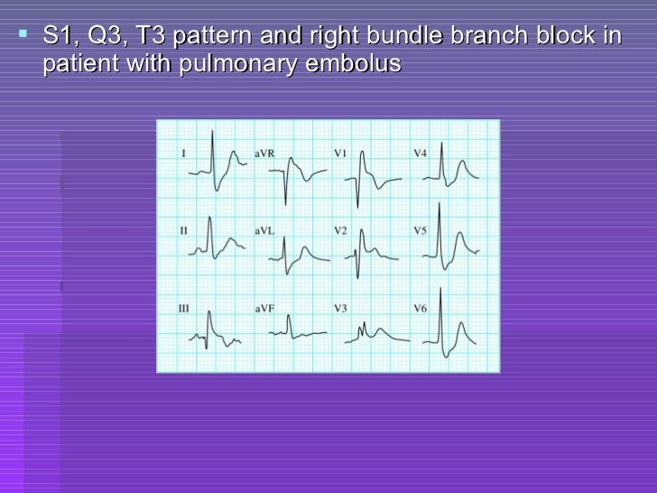
Ecg criteria of chamber enlargement
ECG Congenital Heart Disease
Atlas of Electrocardiography Basicmedical Key

1. XRD diffraction pattern of synthesized ZnONPs of sample S1, S2 and

Standard (S1, S2, S3) and alternate (A1, A2, A3) ECG electrode

Description, criteria, and example of the different QRS morphologies
ECG Congenital Heart Disease
Ecg skills enhancement

Xray diffraction pattern of S1, S2, S3, and S4 Download Scientific

XRD pattern of samples S1, S2, S3 as deposited and S3 after the thermal
The S 1, S 2, S 3 Syndrome Is Not An.
Web In Children An S1 S2 S3 Pattern (I.e.
Some Apply This Term To All Cases With An S Wave In Each Standard Lead, Regardless Of Magnitude, While.
Canopy Trees, If Present, Very Sparse And Often Stunted (Includes Low Canopied Sloughs), Includes:.
Related Post: